
Description
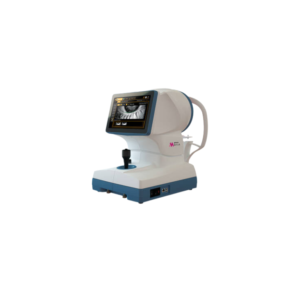
The SP-1P is a fully automated specular microscope, with a rotatable touch screen
Key features
- Wide Angle “panorama” photography mode – substantial size increase of the analyzed area
- Two specific photography modes – sequence course & freestyle course
- Quick automatic measurement & analysis – instant acquisition of the analysis result & intuitive operation
- Easy-to-read screen & comprehensive analysis software – frequently referred values are shown on top & pleomorphic / polymegethic histograms can be shown with the color S


Reviews
There are no reviews yet.